Introduction
In the previous blog, Waterscapes and Where Rivers are Born, we took a peek at the hyporheic zone and saw how this ecosystem lives between the stream’s benthos (bottom) and groundwater. Here we take a closer look at these two worlds as well as a wider view to check out the ecology of groundwater and springs. We’ll see how intricate and widespread this unseen world is and how something ecologists call “connectivity” is crucial to ecosystems.
Lifting the Curtain- Animalcules
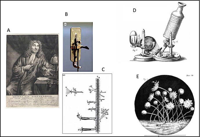
Antoni Van Leuwenhoek’s first microscopic descriptions of his pond Animalcules (1676) and Robert Hook’s revealing 1664 drawings of a wide variety of things he saw in his microscope (he even describes the crystals in urine) (Figure 1.) You may have come across his famous drawing of a flea at some point (Figure 2). His 17th century book Micrographia was wildly popular in scientific circles. It is believed by some to be the first scientific “best seller.” These must have been mystical times, when the biological domain was suddenly growing exponentially!
The Awe and Allure
More impressive views of the Animalcules were revealed in the late 19th century, when microscopes were much better. Even smaller critters (e.g., bacteria) could be seen, and miraculous views of details inside of the Animalcules. The transparent Daphnia (water flea) must have been a star. Even today countless biology students have seen its beating heart and other organs in lab.
I imagine the allure of these critters is even greater today. In Robert Hooke’s time books were dreadfully expensive but now books, images and videos are readily available on the internet. BTW, have you ever tried the Internet Archive? It has countless free books to download or digitally borrow.
Of course microscope technology continues the onward march with both incredible optical and electronic advancements (I recommend checking out the Nikon Small World website for some inspiring images and videos.) Take a peek at the water bear in Figure 8 for an electron microscopic example.
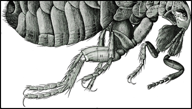
Robert Hooke’s Drawing of a Flea revealed new details (note: just the lower part is shown)
Rotifers, the Wheel Animalcules
I’m always mesmerized by the very hard-working Rotifers in the phylum Rotatoria. They catch their food with what look like spinning circular buzzsaws of synchronized cilia, which is why they were once called “Wheel Animalcules”. They are widespread in freshwaters and moist habitats, from puddles to deep lakes, as well as in semi-moist terrestrial environments like soil and moss. They are real survivors too, reviving after long dry periods for years (in one case in dry moss for 27 years.) They have even survived the harsh liquid traps of carnivorous pitcher plants.
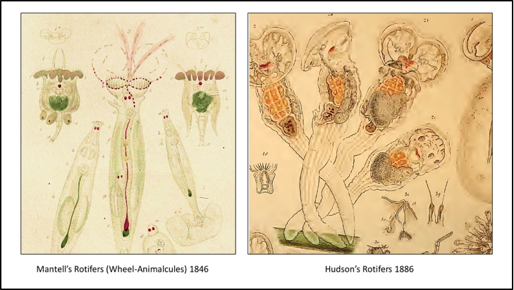
Our views of Rotifers increased significantly with the evolution of better microscopes. The drawings in Figure 3 were 40 years apart, from 1846 to 1886. Mantel’s microscopic drawings revealed amazing detail for the time but Hudson’s drawing in 1886 shows much more detail. You can almost see those cilia creating vortices to pull their food into their “mouths”! I do think they would be great subjects for a remaking of “Alien.”
The Shock
These awesome images also became sources of fear, when these creatures came to be associated with waterborne disease in the 19th century. The Thames River in London was contaminated with sewage and all sorts of obnoxious waste. People were shocked when they saw the microscopic animals and algae in just a drop of their river water. What they did not realize that waterborne diseases like Typhus and Cholera were caused by creatures much smaller than these “animalcules.” Even though they were “not guilty,” their often stylized images did prove useful. Their images benefited a political fight that resulted in massive civil engineering projects to keep wastes out of the Thames River and stop the “big stink.” as it was called.
Left: Rotifer, Pl XII from Mantel’s 1846 book; Right: Part of one of Hudson’s 1886 beautiful and detailed drawings of Rotifers
Groundwater Ecosystems
From Ponds and Streams to Under the Ground
So far we have been talking about critters from ponds, streams and rivers. We now turn to the topic of groundwater because many of those creatures also show up there. Seeing groundwater as an ecosystem is a relatively recent concept, so we start with threats to its existence due to pollution and over pumping.
Baltimore Drank Groundwater and Then It Didn’t
A series of great achievements in public health occurred during the 19th century, born of the need to manage water and sewage to control disease. The golden era of sanitary engineering (late 19th and early 20th century) was crucial to delivering clean water by public works agencies using Science and Engineering (e.g., pipe networks, sewage and water treatment plants.)
People in cities like Baltimore came to realize that human and animal waste in surface waters like streams and lakes was not the only impediment to clean drinking water. The springs and public and private wells were polluted too because of overflowing sewage from cesspools and privies that had soaked into the ground. Baltimore therefore searched for clean water away from the city. Lake Roland was one of these new supplies but it quickly became polluted and the Gunpowder River was selected. The first Loch Raven reservoir dam and tunnel to Lake Montebello and Lake Clifton was built in 1881. Water Engineering has produced incredible systems for the treatment of water, sewage and runoff that serve us well today.
Did you know that even if you don’t use a well, you are still likely drinking groundwater? A lot of the water you see in Baltimore’s reservoir system actually comes from groundwater delivered by its many streams. Those flows are made up of groundwater, not just runoff from storms.
Groundwater is Disappearing
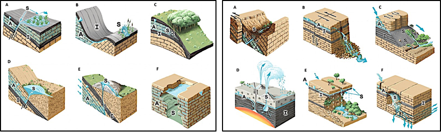
Polluted groundwater can travel great distances, so treatment is rarely feasible on a large scale. Groundwater is a source for some wetlands and many streams, and creates unique terrestrial ecosystems like springs (Figure 4.) The threats to our drinking and irrigation water, as well as groundwater ecosystems continue to increase, due to stresses like unwise water use, urbanization, climate change just to name a few. Konikow (2013) found that 40 US aquifers were depleted by 264 Trillion gallons from 1900–2008 (ca. double all of the water Lake Erie!)
Replenishing groundwater aquifers is often not feasible. For example, the Ogallala aquifer in the American great plains is one of the world’s largest aquifers. It suffers from over pumping and if depleted would take at least 6,000 years to refill (Biello 2012.) Sometimes depletion so severe the land itself drops (subsidence.) Pumping since the 1920’s from thousands of wells in California’s San Joaquin valley has caused the land surface to sink by as much as 28 feet (NASA 2017)!
Groundwater stressors impacts water supplies and surface water ecosystems alike. It is not just streams that are threatened by urbanization, drought and climate change. So are groundwater ecosystems!
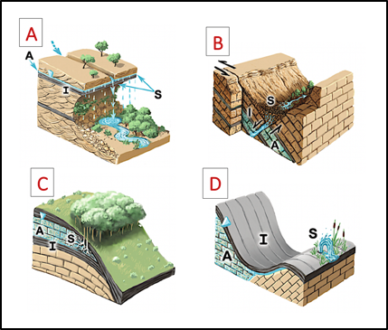
Four Types: A, Hanging Gardens; B, Rheocrene; C, Hypocrene; D, Fountain. Within figures: A, Aquifer; I, Aquitard (barrier); S, Surface Spring; Source: Stevens et al. 2020
Groundwater Ecology
Groundwater and Groundwater Dependent Ecosystems
When groundwater flows are connected to the surface a variety of ecosystems are created like wetlands, springs, seeps, etc. These are called groundwater dependent ecosystems (GDE) and many are unique. GDEs are especially important in arid climates, where they create oases with a rich flora and fauna. These ecosystems are an example of worlds within worlds connected by common water sources.
Interstitial Spaces (aka nooks and crannies)
Most folks don’t realize that groundwater biota live in the nooks and crannies of the subsurface, i.e., the spaces between cobbles, pebbles, sand, sediments and rocks (Figure 5.) Picture water flowing within these interstitial spaces large and small. They seem like little streams and river networks, but the water moves very slowly, as it follows millions of tiny twists and turns. In groundwater, an overall velocity through the aquifer of a foot per day is considered fast; it can also be only a foot per decade!
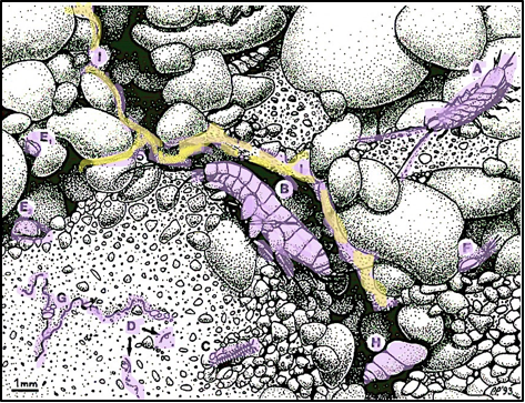
Figure 6 shows a more realistic biological view of these spaces and the aquatic organisms who live there. You can also see biofilms, made of bacteria (the yellow color in Figure 6). The rocks, spaces and critters are real ecosystems, complete with species interactions, food webs, processing of nutrients, etc. It is dark down there, and those bacterial biofilms are an important food for the critters that live there (there is no grass, so grazers have to eat biofilms made of bacteria.) Maybe we can call the scene in Figure 6 the “Star Wars” hotel?
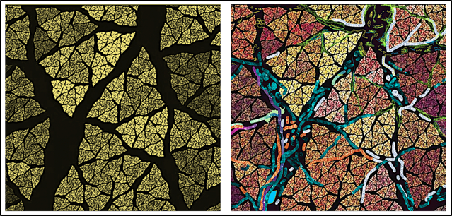
Empty spaces (Left), and with a diversity of microbial and insect Life (Right). Source: Backgrounds from part of a creation by artist Doug Morgan; Stylized microbial life in cracks by KTB
Groundwater biota of interstitial spaces; A, Isopod; B, Amphipod; C, Groundwater Malacostran; D, Copepod; E1 E2, Ostracods; F, Copepod; G, Segmented Worm, H, Gastropod (Snail); I, Bacterial Biofilm. Source: Danielopol et al. 1994.
Leaky Boundaries Create Diverse Ecosystems
It is important to remember that the stream’s benthos, hyporheic and groundwater ecosystems have very leaky “boundaries” (take a sneak-peak at Figure 15). So you might find the same organisms in both the hyporheos and groundwater, while others specialize and stay in the groundwater or the benthos, in the hyporheos. For example the young instars (life stages) of many aquatic insects will live in the hyporheos to escape currents and predators and then move upward when they are large enough to avoid currents and predation. Some stoneflies spend most of their lives in the aquifer next to the river, only returning as adults to lay eggs in the river.
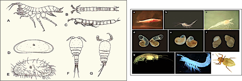
The creatures of the hyporheos and groundwater are diverse (see Figure 7.) Their species, size and numbers tend to change along a vertical gradient as interstitial spaces get smaller and dissolved oxygen values decrease. Specialists and generalists are both part of the community. Ecosystem leakiness facilitates the creation of diverse biota and habitats. There are many ways to make an ecological living!
The Water Bears
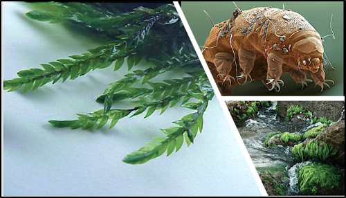
One intriguing critter can be found in the hyporheic zone and in many other very different places- terrestrial moss, aquatic moss, freshwater sediments, groundwater, springs, seeps, etc. They are the Water Bears, or Tardigrades (see Figure 8.) They tend be abundant in lichens and mosses but live in many kinds of environments. They have even been found in activated sludge facilities at sewage treatment plants. Water Bears only need a thin film of water to live in, but that is not available they are famous for entering a protective dehydrated state for very long times. Despite a lifetime of only a few months, they have survived dehydration for up to 30 years. Water Bears can endure a plethora of toxins and environmental stresses that would kill most animals. This is likely part of the reason that they go way back in time; they have survived all five of the Earth’s mass species extinctions!
A sampling of some invertebrates living in groundwater. Left, (A, Amphipod; B,C, Copepod; D/E Ostracods; F/G Copepods) Right, (Fish, Crustaceans, Gastropods (Snails), and an insect. Source: Humphreys et al. 2009.
These are one of the stranger critters of the Hyporheic Zone (Top Right). These little cuties are widespread in many moist environments, like moss (Left, Lower Right). Sources: Left: Britannica.com; Top Right, Micropia; Lower Right, Pxfuel.com
Springs and Groundwater Based Ecosystems
Three Million Springs But Understudied
Springs and seeps are really important because they are places where groundwater “pops up” at the surface. They are of course sources of drinking water but can also have historical meaning and in some cases cultural reverence. Most people don’t realize how diverse springs and their ecosystems can be (see Figure 9.) Though there are more than three million springs on Earth yet they are understudied ecosystems (Stevens et al. 2020.)
Springs- More than Drinking Water
Figures 10 and 11 illustrate some of the ecosystem services associated with springs. The springhouse at the US NPS Hampton Historical Site in Towson must have been indispensable to the estate. Built before 1800, this springhouse provided water and served as a “dairy” where milk was kept to keep it cool (springs can have very cool waters.) The Quitobaquito Spring in Arizona creates a large pool that is important to Native American culture and as a GDE it is an oasis for an ecosystem in a very dry climate. Despite the fact that it is in a National Park, the water levels have dropped to their lowest level in more than a decade, while the spring’s flow rate has reached an all-time low (July 2020.) Nearby urban development and construction have threatened its existence by interrupting nearby groundwater flow patterns.
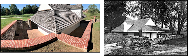
Springhouse at Hampton National Historic Site, Towson MD. Left, Spring House today (KTB photo); and when it was used as a “dairy” to store milk (US NPS photo)

Top: view of Spring; Bottom Left: Dried-up portion of Quitobaquito spring with turtle tracks; Bottom Right: The threatened Sonoran Mud Turtle (lives in this and other springs, rivers); Sources: Top, US National Park Service (USNPS); Bottom, Left: National Geographic; Bottom, Right,Tucson Herpetological Society
Biofilms and Microbiomes
The Structure of Rock Slime
One of our hidden water worlds turns out to be one of the most widespread and consequential to life on Earth- biofilms. As any angler knows, walking on stream rocks can be as slippery as ice (being an avid stream walker I’ve slipped many a time.) This slime is far more than just an algal biofilm. In fact they are intricate, complex ecosystems with a structure that creates protection for its members and that enables the processing of stream nutrients and other chemicals (Figure 12, Left and Upper Right).
Slimy coatings on steam rocks are made of a rich communities of bacteria, fungi, algae, insects and other critters. These biofilms remind me of a forest, with upper and lower canopies and understory vegetation. Different stream critters occupy different heights. Some graze on diatoms and algae on the surface of the rock (e.g., snails and some Mayfly nymphs), while others feed and hunt in the upper canopy (gathering, shredding and piercing modes of feeding.) Think of it as a slime hotel, with different kinds of restaurants on every floor.
Diatom Boats
One of my favorite critters of the slime are the benthic Diatoms (Figure 12, left). I had decided to take a microscopic look at the biofilm of a benthic brick from Stony Run (near JHU) and saw a metropolis of life, with many beautiful creatures. However what caught my microscopic eye were the green Diatoms moving about with no visible means of transportation (like cilia or flagella). They move on a slime trail, analogous to that of snails, and are an important part of the biofilm ecosystem, providing food for insect and snail grazers. Many of the Diatoms are motile and the ones shaped like boats remind me of a harbor full of moving ships, adding to their charm.
Modern Views of Biofilms
Scientists were aware of these biofilms in the 19th century (called Auwfuchs then), but we had to wait for 20th century microscopy to begin to realize their real diversity and structure, about 40 years ago (Figure 12, Bottom Right.) The great revelation was that benthic bacteria were key to their structure and function. Along with algae, protozoans, fungi and tiny invertebrates, they actually govern stream microbial life. Today, they are called Periphyton or Epilithon but I rather like the older “Auwfuchs”, as it sounds more intriguing .Today biofilms are seen as important parts of so many natural and not-so natural ecosystems, including within our guts, the insides of water pipes, as parts of sewage treatment processes, and around soil particles and plant roots, just to name a few. We realy could not survive without them!
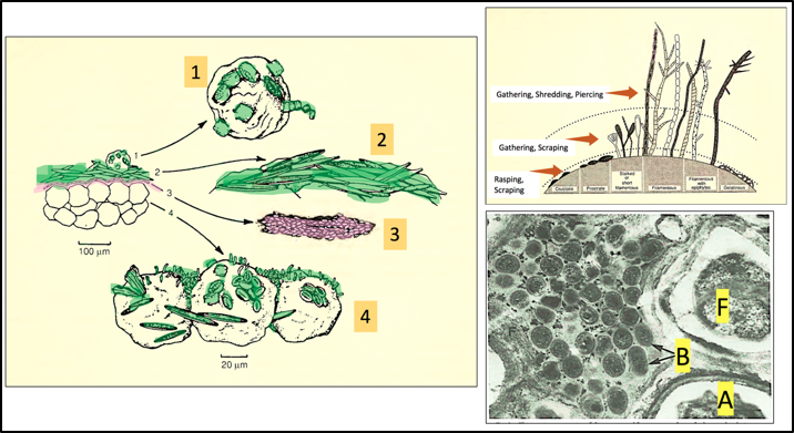
Left: Layers of Periphyton (Aufwuchs) growing on sand grains in a stream. 1) Diatoms on sand grains easily moved by the water current; 2) “Upper story” mats of motile diatoms 3) Mucilaginous layers of diatoms. 4) “Understory” sessile biota (non-motile); Top Right: Various growth forms of Periphyton, showing how these have consumers that focus on particular growth forms (the three zones.); Bottom Right: Thin section of “slime” from benthic rock surfaces in a mountain stream; Bacterial (B) and Algal cells (A) within a matrix of slime fibers (F); Sources: Left and Top: Allan and Castillo 2017, Bottom Right: Geesey et al. 1978.
Biofilms Everywhere
Biofilms are almost everywhere we look in nature, from stream and groundwater systems, plant leaves and roots, and around soil particles (Figure 13, Left and Right, respectively.) As water flows through and around these tiny surfaces, it makes its way to streams and groundwater. Battin et al. (2016) refer to these biofilms as the watersheds’ “microbial skin”.
Biofilms can have a lot to do with improving water quality in groundwater and streams. The hyporheos, with its complex system of water flows and biofilms is good at removing stream pollutants; in fact it has been referred to as the “rivers liver.”
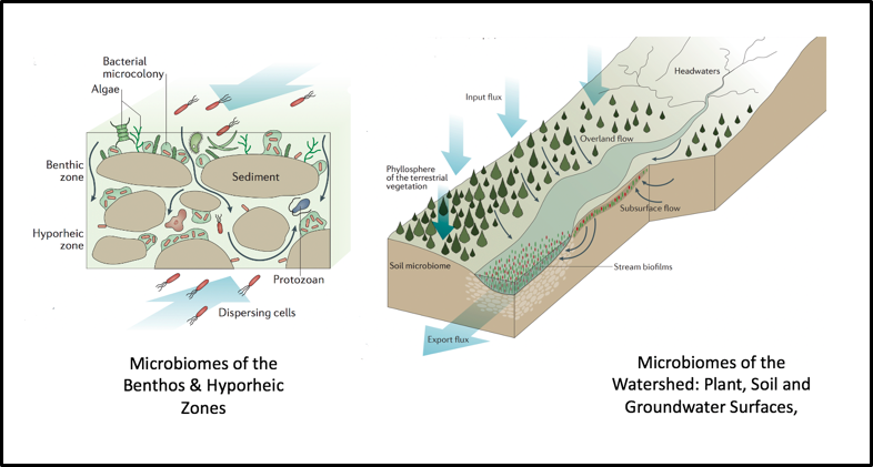
Microbiomes of the benthos & Hyporheic Zone (Left); Right: Expanded watershed-wide microbiomes. Source: Battin et al. 2016.
Interconnected Webs of Water Worlds
Ecosystems within Ecosystems
Let’s look at Figure 14 for a bit. It gives one a feeling for the wonderful complexity of aquatic ecosystems. The benthic and groundwater critters change, depending on the view from different spots in this three dimensional space. Imagine all these microenvironments and the interconnected webs of water, nutrients, chemicals and critters going this way and that. We’ll get back to this figure in a future blog, but for now, it gives us an idea of the wonderful complexity beneath our feet.
Micro, Macro and the Moving Parts
We’ve caught a glimpse of life in various hidden water worlds, from the microscopic to the macroscopic (Figure 15.) and have seen some repeating themes.
First, life is not a haphazard collection of critters. It is organized into communities and ecosystems that have a wide array of habits, structures and functions. Second, we’ve seen that microbes revealed by microscopes have turned out to be very important parts of aquatic ecosystems. Third, aquatic ecosystems are incredibly connected and that boundaries between the benthos, hyporheos and groundwaters are leaky.
It is good to remember that ecosystems are systems! They are like giant machines with lots of moving parts. When we throw a monkey wrench into them they can degrade or even fall apart.
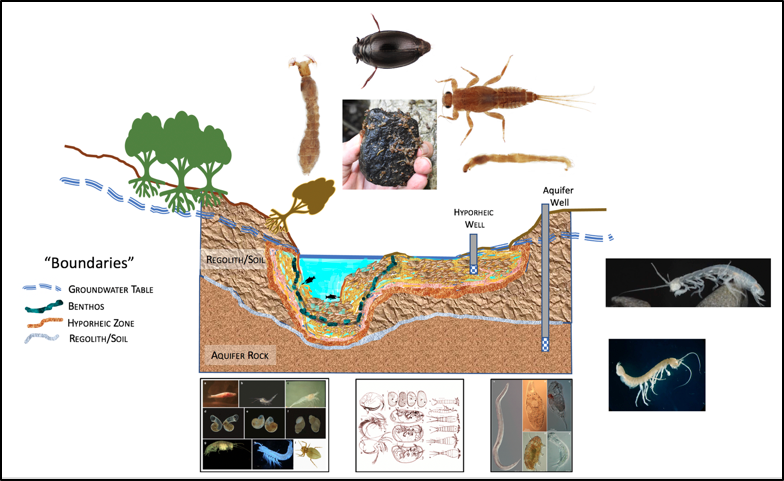
Different kinds of environments with different kinds of critters. The “boundaries” on the left roughly define different environments and ecosystems, as one moves downward. Source; Drawing by KTB (but the critters are from various sources in the bibliography)
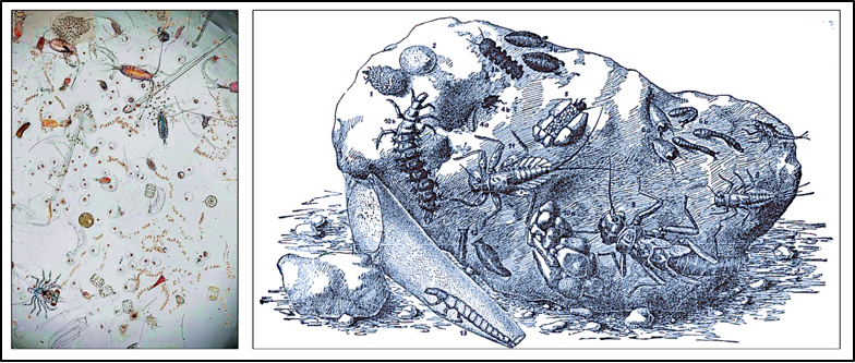
Left, Aquatic Microorganisms; Right, Various Macroinvertebrates Associated with Rocks on the Stream Benthos. Sources: Left, Unknown; Right, Ruttner 1963 (Enhancement by KTB)
The Swirl of Science and Groundwater
The study of groundwater has been the purview of the field of Hydrology (the study of water and its flows) for a long time. Today we know that groundwater is a key part of a number of ecosystems and that understanding these requires a holistic, interdisciplinary approach Take a look at the “word cloud” from a literature search of thousands of scientific articles (Figure 16, Left side.) The search terms “Ecology” and “Hydrogeology” yielded articles that reveal many different, overlapping approaches and issues.
The artwork’s vortices on the right of Figure 16 remind me of the tangle of groundwater flowpaths. They are everywhere, even though we cannot see them. You might imagine these swirls heading into the depths, bouncing off rocks and heading down a maze of tiny tunnels. Can you imagine the critters flowing and twirling in a big “critter hoedown”? How about all that knowledge swirling around those critters!
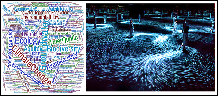
Left: The word cloud illustrates the many topics and water problems that emerge from the disciplines of “Ecology” and “Hydrogeology.” Right: This artwork by Toshiyuki Inoko is reminiscent of how the many vortices of groundwater flow are everywhere.
Bibliography and Credits
Allan, J. D.; Castillo, M. M., Stream ecology: structure and function of running waters. Springer Science & Business Media: 2007.
Britannica: https: //www.britannica.com/plant/water-moss;
Battin, T. J.; Besemer, K.; Bengtsson, M. M.; Romani, A. M.; Packmann, A. I., The ecology and
biogeochemistry of stream biofilms. Nature Reviews Microbiology 2016, 14 (4), 251-263.
Biello, D., Farmers Deplete Fossil Water in World’s Breadbaskets. Scientific American: 2012;
Danielopol, D. L.; des Châtelliers, M. C.; Moeszlacher, F.; Pospisil, P.; Popa, R., 8 – Adaptation of Crustacea to Interstitial Habitats: A Practical Agenda for Ecological Studies. In Groundwater Ecology, Gibert, J.; Danielopol, D. L.; Stanford, J. A., Eds. Academic Press: San Diego, 1994; pp 217-243.
Fischer, H., F. Kloep, S. Wilzcek, and M. T. Pusch. “A River’s Liver – Microbial Processes within the Hyporheic Zone of a Large Lowland River.” Biogeochemistry 76, no. 2 (Nov 2005): 349-71
Geesey, G. G., Mutch, R., Costerton, J. W. & Green, R. B. Sessile bacteria an important component of the microbial population in small mountain stream. Limnol. Oceanogr. 23, 1214–1223 (1978).
F. R. Hauer & G. A. Lamberti 2017 Methods In Stream Ecology, Internet Archive
Hooke, Robert. 1664 Micrographia: Some Physiological Descriptions of Minute Bodies Made by Magnifying Glasses with Observations and Inquiries Thereupon. London: John Martyn, and James Allestry, Printers to the Royal Society of London. Note: Excerpts from Hooke’s famous Micrographia book is available for free on the Project Gutenberg website as an Ebook at www.gutenberg.net
Humphreys, W. F., Hydrogeology and groundwater ecology: Does each inform the other? Hydrogeology Journal 2009, 17 (1), 5-21.
Internet Archive. https://archive.org/
Inoko, Toshiyuki -TeamLab “Moving Creates Vortices and Vortices Create Movement), 2017, by, on view at the National Gallery of Victoria, Melbourne. https://www.wallpaper.com/art/teamlab-vortex-installation-ngv-triennial
Konikow, L. F. Groundwater Depletion in the United States (1900–2008); Scientific Investigations Report 2013−5079; U.S. Department of the Interior, U.S. Geological Survey: Reston, VirginiaU.S. Department of the Interior, U.S. Geological Survey, 2013; p 63.
Mantell, G. A., Thoughts on Animalcules; or a Glimpse of the Invisible Word Revealed by the Microscope. John Murray: London, Albemarle Street, 1846.
Micropia: https://www.micropia.nl/en/discover/microbiology/water-bear/
Moran, Douglas: https://pixels.com/profiles/doug-morgan
NASA San Joaquin is still Sinking. https://earthobservatory.nasa.gov/images/89761/san-joaquin-valley-is-still-sinking (accessed 10/0620).
National Geographic: Sacred Arizona spring drying up as border wall construction continues, Nat.Geogr. July 2020, by D. Main, Photos by A. Ponders https://www.nationalgeographic.com/science/2020/07/quitobaquito-springs-arizona-drying-up-border-wall/
Nikon Small World: https://www.nikonsmallworld.com/
Pxfuel: https://www.pxfuel.com/en/free-photo-xqbdt
Ruttner, F., Fundamentals of Limnology. Univ. Toronto Press, Toronto: 1963.
Stevens, L. E.; Schenk, E. R.; Springer, A. E., Springs ecosystem classification. Ecological Applications 2020, n/a (n/a), e2218.
Tucson Herpetological Society: Sonoran Mud Turtle:; https://tucsonherpsociety.org/amphibians-reptiles/turtles-tortoises/sonora-mud-turtle/
US National Park Service (NPS): https://www.nps.gov/orpi/learn/historyculture/quitobaquito-springs.htm?TB_iframe=true&width=921.6&height=921.6
List of Figures
Figure 1 The Microscopes of Leeuwenhoek and Hooke
Left, A, Antonine Van Leeuwenhoek, B, Leeuwenhoek’s microscope and C, His drawing of Rotifers on a plant stem; Right: D, Robert Hooke’s microscope and E, His drawing of “Blue Mould”
Figure 2 The Flea: Up Close and Personal
Robert Hooke’s Drawing of a Flea revealed new details (note: just the lower part is shown)
Figure 3 Rotifers and Microscope Advancement in the late 19th Century.
Left: Rotifer, Pl XII from Mantel’s 1846 book; Right: Part of one of Hudson’s 1886 beautiful and detailed drawings of Rotifers
Figure 4 Examples of Terrestrial Ecosystems Created by Springs
Four Types: A, Hanging Gardens; B, Rheocrene; C, Hypocrene; D, Fountain. Within figures: A, Aquifer; I, Aquitard (barrier); S, Surface Spring; Source: Stevens et al. 2020
Figure 5 Groundwater “Nooks and Crannies”
Empty spaces (Left), and with a diversity of microbial and insect Life (Right). Source: Backgrounds from part of a creation by artist Doug Morgan; Stylized microbial life in cracks by KTB
Figure 6. Subterranean Critters of the Nooks and Crannies
Groundwater biota of interstitial spaces; A, Isopod; B, Amphipod; C, Groundwater Malacostran; D, Copepod; E1 E2, Ostracods; F, Copepod; G, Segmented Worm, H, Gastropod (Snail); I, Bacterial Biofilm. Source: Danielopol et al. 1994.
Figure 7 Some Groundwater Critters
A sampling of some invertebrates living in groundwater. Left, (A, Amphipod; B,C, Copepod; D/E Ostracods; F/G Copepods) Right, (Fish, Crustaceans, Gastropods (Snails), and an insect. Source: Humphreys et al. 2009.
Figure 8 Water Bears (Tardigrades)
These are one of the stranger critters of the Hyporheic Zone (Top Right). These little cuties are widespread in many moist environments, like moss (Left, Lower Right). Sources: Left: Britannica.com; Top Right, Micropia; Lower Right, Pxfuel.com
Figure 9 Some of the many Kinds of Spring Ecosystems. Left, Lentic (standing water) springs; Right, Lotic (flowing or running water) springs. Source: Stevens et al. 2020
Figure 10 19th Century Springhouse at Hampton Estate
Springhouse at Hampton National Historic Site, Towson MD. Left, Spring House today (KTB photo); and when it was used as a “dairy” to store milk (US NPS photo)
Figure 11 Quitobaquito Spring in Arizona;
Top: view of Spring; Bottom Left: Dried-up portion of Quitobaquito spring with turtle tracks; Bottom Right: The threatened Sonoran Mud Turtle (lives in this and other springs, rivers); Sources: Top, US National Park Service (USNPS); Bottom, Left: National Geographic; Bottom, Right,Tucson Herpetological Society
Figure 12 Benthic Stream Biofilms
Left: Layers of Periphyton (Aufwuchs) growing on sand grains in a stream. 1) Diatoms on sand grains easily moved by the water current; 2) “Upper story” mats of motile diatoms 3) Mucilaginous layers of diatoms. 4) “Understory” sessile biota (non-motile); Top Right: Various growth forms of Periphyton, showing how these have consumers that focus on particular growth forms (the three zones.); Bottom Right: Thin section of “slime” from benthic rock surfaces in a mountain stream; Bacterial (B) and Algal cells (A) within a matrix of slime fibers (F); Sources: Left and Top: Allan and Castillo 2017, Bottom Right: Geesey et al. 1978.
Figure 13 Microbiomes in Streams and Watersheds
Microbiomes of the benthos & Hyporheic Zone (Left); Right: Expanded watershed-wide microbiomes. Source: Battin et al. 2016.
Figure 14 The Stream Biota Waterscape and Leaky Boundaries.
Different kinds of environments with different kinds of critters. The “boundaries” on the left roughly define different environments and ecosystems, as one moves downward. Source; Drawing by KTB (but the critters are from various sources in the bibliography)
Left, Aquatic Microorganisms; Right, Various Macroinvertebrates Associated with Rocks on the Stream Benthos. Sources: Left, Unknown; Right, Ruttner 1963 (Enhancement by KTB)
Left: The word cloud illustrates the many topics and water problems that emerge from the disciplines of “Ecology” and “Hydrogeology.” Right: This artwork by Toshiyuki Inoko is reminiscent of how the many vortices of groundwater flow are everywhere.